
Publication Date
5-2019
Files
Download Full Text (778 KB)
Mentor, Preceptor, Principle Investigator
Mentor: Paul Grabb MD
Abstract
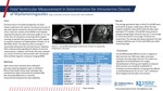
Prenatal closure of myelomeningoceles for fetuses with ventricular width of greater than 15 mm at the time of intrauterine screening (19-25 weeks) has been discouraged, but little is documented regarding the details of ventricle measurement, modality, and timing. This study concludes that ultrasound and MRI provide different results in regards to fetal ventricle size. If treatment recommendations are going to be offered or withheld based on the 15 mm "rule" the method of fetal imaging must be taken into account.
MeSH Keywords
Myelomeningocele; Fetal Therapies; Ultrasonography; Magnetic Resonance Imaging; Comparative Study
Keywords
Fetal Surgery; Ventricle Measurement; Spina Bifida
Disciplines
Congenital, Hereditary, and Neonatal Diseases and Abnormalities | Obstetrics and Gynecology | Pediatrics | Surgery
Recommended Citation
Lundy, Paige; Vlastos, Emanuel; and Grabb, Paul A., "Fetal Ventricular Measurement in Determination for Intrauterine Closure of Myelomeningoceles" (2019). Posters. 108.
https://scholarlyexchange.childrensmercy.org/posters/108
Included in
Congenital, Hereditary, and Neonatal Diseases and Abnormalities Commons, Obstetrics and Gynecology Commons, Pediatrics Commons, Surgery Commons


Notes
Presented at Children's Mercy Kansas City Research Days, May 2019.