
Publication Date
12-2022
Files
Download Full Text (367 KB)
Abstract
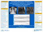
Introduction: Hypereosinophilic syndrome (HES) is a rare but detrimental diagnosis in the pediatric population. Cardiac involvement within HES can present as a diagnosis of Eosinophilic Myocarditis which can progress to irreversible damage and potentially death. This case report discusses a 16-year-old male treated for HES, although HES can have overlap with Eosinophilic Granulomatosis with Polyangiitis (EGPA) and requires a multi-disciplinary approach to patient management. Case Description: The patient was a 16-year-old male, with a history of asthma, presenting with acute onset of chest pain. He was found to have peripheral eosinophilia with an absolute eosinophil count of 11.71. He had ST segment elevations on electrocardiogram, persistently elevated troponin levels (peak of 14.8ng/mL), and increasing arrhythmia burden which consisted of multifocal premature ventricular contractions and rate dependent bundle branch block. A cardiac MRI was notable for an edematous left ventricle with moderate to severe dysfunction, moderate right ventricular dysfunction, and moderate pericardial effusion. Endomyocardial biopsy was not performed given the tenuous clinical status of the patient and he was admitted to the pediatric cardiac intensive care unit. A diagnosis of Eosinophilic Myocarditis was made based on clinical, laboratory, and imaging changes for which high dose steroids were administered. There was a dramatic decrease in peripheral eosinophilia (0.21) as well as near resolution of arrhythmia burden within twenty-four hours of steroid initiation. He successfully discharged from the hospital but continued to have eosinophilia in the outpatient setting. Given the working diagnosis of HES and possibility of EGPA, therapy with mepolizumab, an IL-5 inhibitor to prevent activation and proliferation of eosinophils and B cells, was initiated and is a treatment for both HES and EGPA. Unfortunately, he has now developed chronic left ventricular systolic dysfunction and dilation which is being managed with sacubitril/valsartan, metoprolol, and eplerenone with which he remains asymptomatic. Discussion: The etiology of the eosinophilia was most likely secondary to HES although there remains overlap with EGPA and is difficult to exclude. EGPA is rare in the pediatric population and is largely a tissue diagnosis. Given that the patient had presented on daily steroids, a tissue diagnosis would have likely been inconclusive, however, the diagnosis of EGPA is considered given the patient’s longstanding history of asthma, history of nasal polyposis with sinusitis, and sudden presentation of myocarditis with eosinophilia. Without definitive tissue biopsy and a negative ANCA, this diagnosis is largely based off presumption and will require monitoring his response to treatment. HES can present with myocarditis and is still the likely etiology, however, the patient had a normal bone marrow biopsy and did not have any gene mutations known to have an association with the disease (JAK2, V617F, and c-kit D816V). Although initial treatment may be similar, the management for refractory HES and EGPA differs and will require ongoing multi-disciplinary follow up.
Disciplines
Critical Care | Pediatrics
Recommended Citation
LaVoy, Nathan; Birnbaum, Brian; Harris, Julia G.; Pandya, Aarti; and Taber, Allison, "Eosinophilic Myocarditis Secondary to Hypereosinophilic Syndrome vs. Eosinophilic Granulomatosis with Polyangiitis: A Diagnostic Dilemma" (2022). Posters. 307.
https://scholarlyexchange.childrensmercy.org/posters/307


Notes
Presented at The Pediatric Cardiac Intensive Care Society Annual Meeting; Miami, FL; December 15-18, 2022.