
Publication Date
5-2022
Files
Download Full Text (1.5 MB)
Abstract
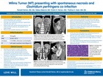
Wilm’s Tumor (WT) is the most common renal malignancy of childhood. The initial presentation of WT may mimic infection or other malignancies with nonspecific symptoms including fever, abdominal/flank pain, or hematuria. We describe a case of WT with spontaneous necrosis obscured by overlying Clostridium perfringens infection. A previously healthy 5-year-old girl presented to the emergency department with a two-day history of flank pain and fevers. She was tachycardic (130 bpm), hypertensive (116/75), and febrile (101.7F). Her abdomen was distended with left-sided flank tenderness without a palpable mass. Initial laboratory testing revealed elevated CRP (70mg/dL) and leukocytosis (22.56 x 10⁹ /L). Urinalysis showed 5-10 WBCs but was otherwise normal. A CT abdomen/pelvis with contrast demonstrated a mass-like structure in the left upper quadrant, surrounding and splaying the left adrenal gland with inferior displacement of the left kidney. The patient was hospitalized for evaluation and received empiric ceftriaxone. Additional laboratory studies were obtained including urine cultures, plasma normetanephrine and metanephrine levels, as well as Vanillylmandelic Acid (VMA) and Homovanillic acid (HVA) levels. All were within normal limits. Her symptoms, leukocytosis and CRP improved with resolution of fevers after 5 days. Given the ambiguous clinical picture favoring infectious versus oncologic diagnoses, open surgical biopsy of the left kidney and surrounding mass was performed. Pathology demonstrated a phlegmonous appearance of the left kidney and fibrovascular proliferation with inflammation and no evidence of malignancy. Tissue cultures were positive for Clostridium perfringens. At discharge, the patient transitioned to a 10-week course of cefixime for C. perfringens kidney infection with outpatient follow-up. Repeat CT abdomen/pelvis obtained 4 weeks after discharge demonstrated decreased size of the renal mass. In outpatient follow-up, repeat ultrasounds minimally improved, therefore a second renal biopsy was obtained 13 weeks after discharge. The biopsy identified monomorphic small cells in varying stages of cell death with positive WT1 protein, consistent with WT. This case features an unusual presentation of WT with spontaneous necrosis and concomitant C. perfringens infection. Our patient’s presentation was suspicious, however her initial negative biopsy was masked by the presence of secondary bacterial infection and necrosis, leading to delayed oncology referral and treatment. This case underscores the importance of avoiding confirmation bias in the setting of ambiguous clinical presentations. WT may mimic other pediatric renal pathologies clinically and radiologically, such as renal hemorrhage, abscess, or other malignancies. This poses a diagnostic challenge for providers, particularly when both infection and malignancy are observed.
Disciplines
Pediatrics
Recommended Citation
Palmen, Ronald; Elsbernd, Abbey; Palmen, Kristin; and Kyler, Kathyrn, "Wilms Tumor (WT) presenting with spontaneous necrosis and Clostridium perfringens co-infection" (2022). Posters. 273.
https://scholarlyexchange.childrensmercy.org/posters/273


Notes
Presented at the American Society of Pediatric Hematology/Oncology (ASPHO) Annual Meeting; Pittsburgh, PA; May 6, 2022