
Publication Date
2-2020
Files
Download Full Text (451 KB)
Abstract
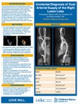
Description of Clinical Presentation: A 15 year old male with a 2 week history of fever and abdominal pain had an abdominal/pelvic MRI demonstrating osteomyelitis of his right hip. Incidentally an anomalous vessel arising from the abdominal aorta and coursing to the vasculature of the lower right lung lobe was identified and he was referred to cardiology for further evaluation. He is an athlete with no history of cardiac or respiratory symptoms and his cardiac physical examination was within normal limits. Diagnostic Techniques and Their Most Important Findings: An echocardiogram was initially obtained; imaging was difficult due to suboptimal acoustic windows and revealed normal intracardiac anatomy and normal ventricular size and systolic function. Detailed profile of the right pulmonary venous connections was not feasible and an abnormal flow close to the inferior vena directed towards the thoracic cavity was seen. The suspicion of an anomaly involving the right lung vasculature was raised and a cardiac MRI (CMRI) was obtained in order to better assess cardiovascular anatomy and calculate Qp:Qs. CMRI revealed an anomalous systemic arterial supply of a small portion of the right lower lobe without pulmonary sequestration. The artery originated from the celiac trunk with proximal narrowing of the vessel close to its origin and post-stenotic dilation. There was normal right pulmonary artery arborization and supply to this segment, indicating dual arterial supply. Qp:Qs was normal (1.03) with normal atrial and ventricular volumes indicating minimal extracardiac shunt. Learning Points from this Case: Incidental MR imaging findings are not uncommon and further imaging work-up as well as management should be individualized. In this case a systemic arterial supply to the right lower lung was inadvertently identified and further imaging revealed no hemodynamic burden associated with it. Various case reports address similar incidental discoveries, usually associated with work-up for hemoptysis, congenital heart disease and often associated with lung sequestration. Ligation or embolization of the systemic feeding vessel to the lung due to risk of hemoptysis is often undertaken. In the case of our patient, the morphology of the feeding vessel in combination with normal lung appearance, normal Qp:Qs, and lack of symptoms led to a consensus to not intervene but to observe with regular follow-up.
Disciplines
Cardiology | Pediatrics
Recommended Citation
Mathis, Christopher; Crockett, Jay; and Kiaffas, Maria, "Incidental Diagnosis of Dual Arterial Supply of the Right Lower Lobe" (2020). Posters. 158.
https://scholarlyexchange.childrensmercy.org/posters/158


Notes
Presented at Society of Cardiovascular Magnetic Resonance 23rd Annual Scientific Sessions. Orlando, Florida.