
Publication Date
6-2021
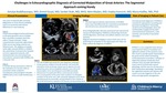
Files
Download Full Text (452 KB)
Abstract
Clinical Presentation
Two cases of {S,D,L} anatomically corrected malposition of great arteries (ACMGA) are presented with differences in conal anatomy. Case 1: A fetal echocardiogram (echo) performed at 28 weeks gestation due to multiple anomalies revealed atrial situs solitus, D-loop ventricles, a large conoventricular septal defect (VSD) and an overriding, anterior and leftward aorta. The diagnosis of double outlet right ventricle (DORV) vs ACGMA was entertained. Postnatal echo revealed {S,D,L} segmental anatomy with ventriculoarterial concordance consistent with ACGMA and a muscular VSD. Case 2: A 3-week-old boy presented to clinic for a murmur evaluation with no associated cardiac symptoms. Transthoracic echocardiogram revealed {S,D,L} ACGMA and a small membranous VSD.
Imaging Findings
Echocardiographic subcostal, parasternal and suprasternal sweeps help diagnose ACGMA by delineating ventriculoarterial alignment, ventricular and outflow relations and conal anatomy. Case 1, a rare ACMGA type, posed the most diagnostic challenges. A more anterior rotation of the left ventricle, horizontal orientation of the ventricular septum due to absence of a sub-pulmonary conus, in combination with an elongated subaortic conus led to the misconception of an overriding aorta and DORV prenatally. Pulmonary to tricuspid valve fibrous continuity was present. The unusual position of the aortic valve resulted in an elongated curvature of a left aortic arch, coursing from anterior and right towards the left of the trachea. Case 2, the most typical type of {S,D,L} ACGM, had the usual anatomic characteristics of bilateral sub-arterial conus and parallel outflow tracts. In both cases cardiac magnetic resonance imaging was performed for evaluation of the Qp/Qs caused by the VSD and confirmed the diagnosis of ACGMA.
Roles of Imaging in Patient Care
Accurate prenatal diagnosis of ACMGA is essential for appropriate counseling and postnatal management. Postnatally, standard echocardiographic views and multimodality imaging will elucidate ventriculoarterial connections, conal anatomy, and severity of associated anomalies.
Discussion
ACMGA is a rare congenital heart disease occurring from failure of involution and rightward rotation of the subaortic conus resulting to a parallel spatial relationship of the great arteries while maintaining ventriculo-arterial concordance. Subtypes include {S,D,L} (most common, usually with bilateral conus) and {I,L,D} with normal, and {S,L,D} and {I,D,L} with transposition physiology. Identifying the segmental anatomy and relations will result in accurate diagnosis of this rare entity.
Disciplines
Cardiology | Pediatrics
Recommended Citation
Buddhavarapu, Amulya; Goyal, Anmol; Shah, Sanket; Madan, Nitin; Hancock, Hayley S.; and Kiaffas, Maria, "Challenges in Echocardiographic Diagnosis of Corrected Malposition of Great Arteries: The Segmental Approach coming Handy" (2021). Posters. 220.
https://scholarlyexchange.childrensmercy.org/posters/220


Notes
Presented at the 32nd Annual American Society of Echocardiography Scientific Sessions Virtual Conference, June 18-21, 2021.