
Publication Date
10-2020
Files
Download Full Text (276 KB)
Abstract
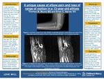
A 13-year-old right hand dominant young lady presented to a pediatric sports medicine clinic for evaluation of decreased ROM and pain in her left elbow with an associated locking sensation that had been occurring for over a year. Two weeks prior to presentation, these symptoms had been acutely worsening as she was practicing for over 10 hours per day for an upcoming dance competition. The patient’s pain is described as a “tightness” and is localized to her antecubital fossa medial to her biceps tendon. The feeling of tightness occurs most prominently with end extension and flexion of the elbow. She has had no associated fever, chills, fatigue, weight loss, swelling, bruising, numbness, tingling, previous reported injury in her elbow, or other pain or mechanical symptoms in any other joint. On exam the patient lacked 5-10° of full extension of the elbow on the left while exhibiting 5° of hyperextension on the right. She is tender to palpation (TTP) over the proximal ulna, flexor tendon mass, capitellum, and biceps tendon as it crosses the antecubital fossa. She is not TTP over the medial epicondyle, lateral epicondyle, olecranon, distal ulna, or on any region of the radius. Initial differential remained broad and included biceps tendinopathy, osteochondritis dissecans, subluxation, dislocation, occult fracture, osteochondroma, osteoarthritis, inflammatory arthritis, and heterotrophic ossification, among other possible etiologies. Frontal, oblique and lateral views of the left elbow showed multiple small heterogeneous calcifications in the antecubital space (the largest measuring approximately 1 cm in greatest diameter). No joint effusion, fracture, or dislocation were present. Soft tissue calcifications are caused by a wide range of pathologies, which can be grouped broadly into dystrophic, iatrogenic, metabolic, connective tissue disease, metastatic, and idiopathic causes. To further elucidate these calcifications an MRI with and without contrast of the left elbow was obtained. The MRI showed a Venous Malformation (VM) with associated phleboliths located anterior to the distal humeral diaphysis, in the region of the posterior aspect of the brachialis muscle, with an associated small intraosseous component in the humerus. VMs are present at birth and grow proportionately as the child develops. They are typically cutaneous, subcutaneous, or mucosal but can be located anywhere throughout the body. VM symptom presentation is highly variable and will often become symptomatic later in life. Treatment is controversial and options include symptomatic support (compression or NSAIDS), surgery, sclerotherapy, and possibly targeted therapy with sirolimus. In this instance the patient was referred to orthopedic surgery and vascular malformation clinic for further management. This case showed Venous Malformation to be a rare cause of elbow pain with associated decreased ROM and a locking sensation in a 13-year-old female patient.
Disciplines
Orthopedics | Pediatrics
Recommended Citation
Munro, Thomas and Harvey, Brian S., "A unique cause of elbow pain and loss of range of motion in a 13-year-old" (2020). Posters. 289.
https://scholarlyexchange.childrensmercy.org/posters/289


Notes
Presented at the AAP National Conference; Virtual; October 2-5, 2020.