
Publication Date
3-2023
Files
Download Full Text (485 KB)
Abstract
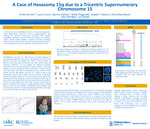
Background: A 7-month-old male with a history of developmental delay, plagiocephaly, hypotonia, chronic cough/congestion was admitted for abnormal movements. Prolonged EEG revealed focal epilepsy and epileptic spasms. Genetic testing revealed a complex structurally rearranged chromosome 15 which contains two inverted duplicated chromosome 15s joined together at one end, resulting in partial hexasomy for 15q. Case presentation: The proband was born to a G2P2 33-year-old mother following an uncomplicated pregnancy at 40 weeks 2 days gestation. At birth he was 6lbs 8oz, 20in long, and APGARs were 3/5/9 at 1/5/10 minutes. At delivery he was limp, pale and had poor tone with minimal crying and respiratory depression. He was admitted due to persistent seizure-like activity. Aa prolonged EEG was abnormal, but movements were not seizures, and he was discharged without medication. A newborn hearing screen was failed, and follow-up confirmatory testing showed mixed hearing loss with right greater than left. At 7-months-of age he was readmitted for seizure. Repeat EEG was indicative of clinical and subclinical focal seizures and epileptic spasms. At his last evaluation at, 11 months of age, he has global developmental delay, hypotonia, and wears bilateral hearing aids. He is unable to sit unsupported but does have head control, is able to roll over and grabs objects with both hands. Imaging studies to date have been negative, including MRI, echocardiogram, and renal ultrasound. Epilepsy is currently well controlled with medication. Microarray testing was ordered at 7 months and showed six copies of an ~7.6 Mb segment between the common breakpoints BP1 and BP4, followed by four copies of an ~1.5 Mb segment between BP4 and BP5 in proximal 15q from 15q11.2 to 15q13.3. Methylation and copy number analysis of 15q11.2q13.1 for Prader-Willi (PWS) suggested maternal inheritance. Chromosome analysis demonstrated a male karyotype with 47 chromosomes including an extra tricentric chromosome. Interphase FISH analysis shows six copies of SNRPN (15q11.2) in 92% of nuclei and two copies of SNRPN in 8% of nuclei. Metaphase FISH analysis found two enhanced SNRPN (15q11.2) signals consistent with a total of four copies of SNRPN in this additional derivative chromosome, indicating the chromosome 15 is composed of two inverted duplicated 15s linked each other at the ends as a way of mirror image. Parental chromosomes were normal, confirming de novo inheritance. Conclusions: Although supernumerary marker chromosome (SMC) 15 itself is common, occurring in ~1/30,000 births, individuals with a tricentric der(15), resulting in partial hexasomy 15q are rarely reported. Complimentary techniques including microarray, MLPA, FISH, and G-banding were used to resolve the structure of the SMC 15. In the future, novel technologies, such as optical mapping, may also be beneficial in the resolution of complex structural variants.
Disciplines
Medical Genetics | Pediatrics
Recommended Citation
Farrow, Emily; Cross, Laura A.; Sullivan, Bonnie; Fitzgerald, Keely M.; Alaimo, Joseph; Repnikova, Elena; Herriges, John; and Zhang, Lei, "A Case of Hexasomy 15q due to a Tricentric Supernumerary Chromosome 15" (2023). Posters. 313.
https://scholarlyexchange.childrensmercy.org/posters/313


Notes
Presented at the Annual Clinical Genetics Meeting; Salt Lake City, UT; March 14-18, 2023.