
Publication Date
4-2023
Files
Download Full Text (304 KB)
Abstract
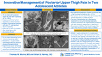
Case history: Two 16-year-old males with posterior upper leg pain. Athlete A reports he had been exploding up to dunk a basketball when he felt a pop with immediate pain in his posterior LLE. He was treated conservatively with activity modification and PT resulting in improvement of symptoms and return to full activity. He returned 15-months after the initial injury due to recurrence of pain. Athlete B stated he had tweaked his hamstring multiple times over the summer and had been working with a physical therapist for rehabilitation. He had attempted full rest for a couple weeks with graded return to activity. He had return to full activity before experiencing a repeat event of severe pain in his posterior upper leg while running at full speed in football. Athlete B stated that if felt like his leg suddenly gave out on him and lost strength. He denies hearing a specific pop. Physical Exam: Athlete A and Athlete B both have normal appearing skin and musculature of the posterior thigh. Both athletes have limited passive extension of the LLE and decreased LLE strength, most significant with resisted knee flexion. Athlete A and B both have focal pain over the left ischial tuberosity with athlete B reporting tenderness extending slightly into the proximal hamstring. No pain is reported when palpating over the ASIS, AIIS, pelvic crest, or greater trochanter. Both athletes have a negative FABER, FABER, and Log Roll. Athlete A has been using crutches to assist with ambulation. Athlete B has a slightly antalgic walking pattern with reported discomfort/pain in left upper extremity. Differential diagnosis: 1) Proximal hamstring strain 2) Ischial tuberosity avulsion fracture/apophysitis 3) Piriformis syndrome 4) Stress fracture (Ischial or femoral) 5) Ischial femoral impingement syndrome Tests and results: - Athlete A: Xray: Left ischial apophysis avulsion fracture. CT: redemonstration of an apophyseal avulsion fracture (1.5cm displaced) with interval development of calcified callus/heterotopic ossification. CT guided fenestration. - Athlete B: Xray: asymmetric appearance the ischial apophyses, with widening/irregularity on the left. MRI Pelvis: Inflammatory marrow signal of the left ischial apophysis. Mild edema around the proximal hamstring tendons. CT guided fenestration. Final/Working Diagnosis: Athlete A: Chronic avulsion fracture of the ischial tuberosity with subsequent nonunion. Athlete B: Acute on chronic ischial tuberosity avulsion fracture. Discussion: There is controversary over the best treatment strategies for pelvic avulsion fractures. In general, non-operative treatment of fractures with < 2cm displacement ( < 1.5 cm for ischial fractures) have a high success rate resulting in healing about 97% of the time. Non-unions are rare, but if they are to occur the ischial tuberosity is a likely location. The use of fenestration as a therapeutic treatment for non-union ischial tuberosity avulsion fractures has been exhibited in a 3 patient case series and 1 case report. We present two athletes with a chronic/acute on chronic ischial tuberosity avulsion fractures who underwent fenestration of the enthesis of the proximal hamstring. Outcomes: Athlete A had initial successful management with pain improvement, healing changes on imaging, and return to sport. Over a 15-month period he exhibited intermittent pain with worsening acute pain. He subsequently underwent an IR guided fenestration. His pain improved and he demonstrated osseous bridging on imaging. Athlete B also underwent fenestration with concerns of acute on chronic injury.
Disciplines
Orthopedics | Pediatrics | Sports Medicine
Recommended Citation
Munro, Thomas and Harvey, Brian, "Innovative Management of Posterior Upper Thigh Pain In Two Adolescent Athletes" (2023). Posters. 318.
https://scholarlyexchange.childrensmercy.org/posters/318


Notes
Presented at the American Medical Society for Sports Medicine (AMSM) 2023 Annual Meeting; Phoenix, AZ; April 27-May 3, 2023.