
Publication Date
9-2025
Files
Download Full Text (456 KB)
Abstract
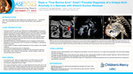
Introduction: The origin of all head and neck vessels from a single aortic root – “True Bovine Arch” - is an extremely rare arch anomaly that has been rarely reported in literature. Clinical Presentation: A 28-year-old G2P1 woman was referred at 27-weeks-gestation for concerns of absent ductus venosus and suspected common atrioventricular valve defect. Fetal echocardiogram revealed bilateral Superior Vena Cava (SVC), dominant left SVC, absent ductus venosus, moderate sized posterior muscular Ventricular Septal Defect (VSD), biventricular dilation with preserved systolic function and a unique arch anomaly with all head and neck vessels arising from a single trunk off the aorta. Postnatal transthoracic echocardiography and CT angiogram confirmed the diagnosis of a true bovine arch with no concerns for coarctation or persistence of a 5th aortic arch. Further systemic imaging revealed dysgenesis of corpus callosum and butterfly vertebrae. A whole exome sequencing was non-diagnostic. Serial follow up echocardiograms revealed the VSD had become restrictive and smaller in size, however, there has been a serial increase in aortic root and ascending aorta dimension. Imaging Findings: Figure 1a shows a sagittal view of the aortic arch with unique branching pattern of all head and neck vessels arising from a single aortic root. Figure 1b and 1c shows post-natal confirmation of the unique aortic arch anomaly via TTE and CTA respectively. Role of Imaging in Patient Care: Accurate prenatal identification of cardiac and aortic arch anomalies is paramount for prenatal counseling, genetic diagnosis, delivery planning and postnatal management. CTA can complement pre and postnatal suspicion for appropriate surgical and endovascular procedural planning. Discussion: The origin of all head and neck vessels from a single aortic root is an extremely rare arch anomaly that has been seldom reported in literature with no documented prenatal identification. A “true bovine arch” refers to a single arterial trunk off the aortic arch that trifurcates into both subclavian arteries and a common bi-carotid trunk—mirroring the configuration found in cattle. Although a common brachiocephalic trunk is considered a normal variant, it has been shown to have a higher likelihood of developing ascending aortic aneurysm and embolic cerebrovascular accidents, a possibility even more likely to happen in our patient with unique arch branching. This could be secondary to hemodynamic variations and sheer stress from an altered angle of branching of the great vessels making identification of these variations vital for risk assessment prior to procedures. Accurate pre-natal delineation of the head and neck vessels can aid in appropriate prenatal counseling, genetic diagnosis, delivery planning and postnatal management especially when associated with critical aortic abnormalities. CT angiogram can complement pre and postnatal suspicion for appropriate surveillance guidelines and pre-procedural planning for surgical and endovascular procedures.
Disciplines
Cardiology | Pediatrics
Recommended Citation
Schermerhorn, Jenna; Abdul Ghayum, Mohamed Aashiq; Kuzava, Laura; Shah, Sanket; and Goyal, Anmol, "Does a “True Bovine Arch” Exist? Prenatal Diagnosis of a Unique Arch Anomaly in a Neonate with Absent Ductus Venosus" (2025). Posters. 476.
https://scholarlyexchange.childrensmercy.org/posters/476
Restricted
This document is restricted to only CMKC staff. Sign in using a CMKC email to access the full text.


Notes
Presented at the American Society of Echocardiography (ASE) 36th Annual Scientific Sessions; Nashville, TN; September 5-7, 2025.