
Files
Download Full Text (627 KB)
Publication Date
5-2022
Abstract
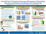
Background: The delta variant of SARS-CoV-2 is reported to cause severe acute lung injury (ALI) in neonates. Neonates with COVID-19 can exhibit a hyper-inflammatory response with vascular injury. The mechanisms by which SARS-CoV-2 structural proteins induce ALI in neonates is unclear. Emerging data suggest that the envelope (E protein) of the SARS-CoV-2 activates Toll Like Receptor (TLR)-mediated immune hyperactivation and cytokine storm. We tested the hypothesis that SARS-CoV-2 E protein can induce TLR-mediated inflammation and ALI in a neonatal mouse model of ALI we developed.
Objectives: 1) To investigate the pathogenicity of E protein in causing neonatal ALI and alveolar remodeling and, 2) To determine whether E protein induces TLR-dependent cell death and inflammation in human pulmonary microvascular endothelial cells (HPMEC).
Methods: We developed a model of systemic E protein-induced ALI using intraperitoneal injections of 10μg (2mg/kg/dose) in P5 [saccular stage] C57BL6 mice. Lung homogenates were used for quantifying cytokine RNA expression and TLR signaling (immunoblotting) 48h after E protein injections. Bronchoalveolar lavage (BAL) was done on P11 mice 48h after i.p E protein. E protein-induced alveolar remodeling was examined by morphometry on P14 after injecting one dose (P5) or two i.p doses (P5 and P8). Neonatal HPMEC (ScienCell) were treated with 500ng/mL E protein, with or without silencing of TLR2 (siTLR2), and cell lysates were examined for cytokine expression and TLR signaling.
Results: E protein induced lung Cxcl1, iNOS, Il-1β, and Tnfαexpression in a dose-dependent manner at 48h. Activation of TLR signaling was evidenced by phosphorylation of inhibitor of kappa B kinase β (pIKKβ) and the NFKB subunit p65 (Fig.1). BAL showed increased albumin content and neutrophil and lymphocyte infiltration 48h after i.p E protein. E protein-induced alveolar simplification was evidenced by decreased radial alveolar counts on P14. (Fig. 2). E protein dose-dependently induced HPMEC cell death [Fig. 3a], as well as TLR signaling [p-IKKβ], NFKB activation [p-p65], and MAPK signaling [p-p38] in HPMEC. E protein (500ng/ml) induced ICAM1, IL-1β, TNFα, IL6, IL8, and TLR2 expression was suppressed by silencing TLR2 using siRNA. (Fig. 3)
Conclusion: The SARS-CoV-2 E protein induces TLR signaling, ALI and alveolar remodeling in the neonatal lung. Ongoing studies suggest that E protein induces direct EC injury in human lung EC that is TLR2-dependent. This study provides mechanistic insight into neonatal immune activation and lung injury seen in infants with COVID-19.
Document Type
Poster
Recommended Citation
Menden, Heather; Yu, Wei; Mabry, Sherry M.; Venkatraman, Aparna; and Sampath, Venkatesh, "The Sars-Cov-2 E Protein Induces Pro-Inflammatory Tlr Signaling, Lung Injury And Alveolar Remodeling In The Neonatal Lung" (2022). Research at Children's Mercy Month 2022. 25.
https://scholarlyexchange.childrensmercy.org/research_month2022/25


